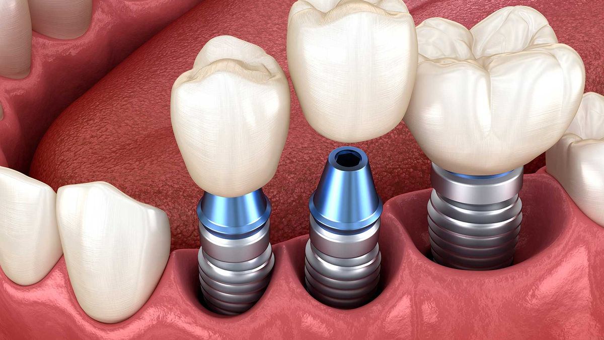
Digital planning means combining images from different sources on a computer to plan implant treatment in advance. This approach helps decide the implant's position based on both the bone structure and the ideal position of the prosthesis to be made.
Digital tools used
Digital photos
Facial and intraoral photos are used for aesthetic assessment, especially in the front of the mouth, and for communicating with the patient.
Digital X-rays
Panoramic and periapical X-rays show the general condition of the teeth and bone.
Cone beam computed tomography (CBCT)
Provides a three-dimensional image of the bone; the position of structures such as the nerve canal, the sinus and neighbouring roots is assessed.
Intraoral digital scan
Creates a surface model of the teeth and gums. When combined with CT data, bone and soft tissue can be seen in the same view.
Planning software
The implant's size, angle and depth are planned virtually; the position of the prosthesis is included in the plan as well.
Surgical guides
Custom-made guides used to transfer the planned position to the mouth. In suitable cases they can help make placement more predictable.
Possible benefits of digital planning
- Detailed assessment of the bone and important anatomical structures
- Prosthesis-driven implant positioning
- Anticipating additional procedures (bone grafting, etc.) in advance
- Sharing the plan visually with the patient
Limits
- A digital plan is a guide; changes may be needed during surgery depending on clinical conditions.
- Guided surgery is not necessary in every case.
- The need for additional imaging is assessed according to the person's situation.
Shared digital planning with orthodontics
Digital models of the teeth are also used in clear aligner treatment. For people with a missing tooth who also need orthodontic treatment, doing orthodontic and implant planning from the same digital data can make the process easier. For details, see our article on clear aligners and implants.


